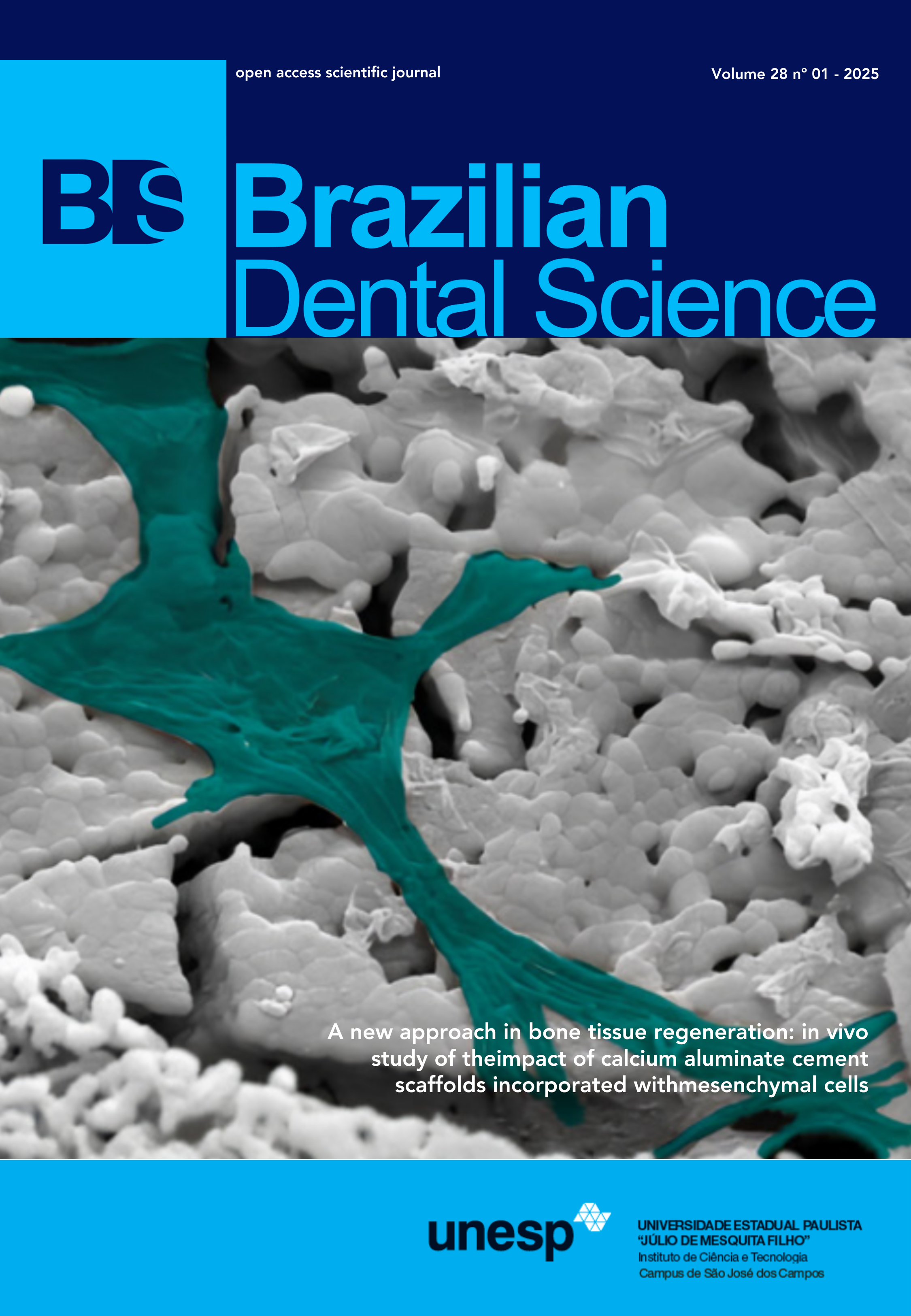
A new approach in bone tissue regeneration: in vivo study of the impact of calcium aluminate cement scaffolds incorporated with mesenchymal cells
DOI:
https://doi.org/10.4322/bds.2025.e4296Abstract
Objective: The objective of this study was to evaluate the bone regeneration potential of CAC-based scaffolds, with or without mesenchymal stem cells (MSC), in bone defects created in rat femurs. Material and Methods: Forty-eight CAC scaffolds and their blends of tricalcium phosphate (TCP), zinc oxide (ZNO), and zirconia (ZIRC) were produced, with half of these incorporated with MSC. Twenty-three Wistar rats were used, with 3 for MSC isolation and 20 for creating bone defects in both femurs. Five animals were assigned to each group, and during the defect surgery and material insertion, the animals received MSC-incorporated scaffolds on the left side and non-incorporated scaffolds on the right side, with the same type of material used in each animal to avoid different systemic effects (n=5); they were euthanized 21 days after the surgical procedure. Results: In the scanning electron microscopy analysis of the scaffolds, structures with open and interconnected pores, as well as cell adhesion, were observed in all groups. In the histological analysis, all groups showed newly formed bone trabeculae interspersed with bone marrow cells and connective tissue. Conclusion: In the histomorphometry, for the scaffolds not incorporated with MSC, the ZIRC group showed greater bone formation, and in the MSCincorporated scaffolds, the TCP group demonstrated better results, both exhibiting a statistically significant difference from the other groups (p<0.05).
KEYWORDS
Biocompatible materials; Bone cements; Bone regeneration; Bone tissue; Mesenchymal stem cells.
Downloads
Published
How to Cite
Issue
Section
License
Copyright (c) 2025 Brazilian Dental Science

This work is licensed under a Creative Commons Attribution 4.0 International License.
Brazilian Dental Science uses the Creative Commons (CC-BY 4.0) license, thus preserving the integrity of articles in an open access environment. The journal allows the author to retain publishing rights without restrictions.
=================


